Domains

____Prokaryotes___

The Eubacteria of Kingdom Monera, commonly called bacteria:
1. Prokaryotic organisms without a nuclear membrane
2. Eubacterial rRNA and no introns in genome
3. Membrane lipids are primarily diacyl glycerol ethers
4. Cell walls contain thick (gram+) or thin layers of peptidoglycan (gram-)

The Archaea, or Archaeobacteria
1. Prokaryotic organisms without a nuclear membrane
2. Archaeobacterial rRNA – introns in genome
3. Membrane lipids are unusual – primarily isoprenoid glycerol diether or diglycerol tetraether derivatives
4. The cell wall does not contain peptidoglycan (gram-)

There are three groups of Archaea, which also called extremophiles: Methanogens are poisoned by O2, Thermophiles live in extreme temperatures, and Halophiles live in highly saline environments

____Eukaryotes___

The Eukarya
1. Nuclear membrane
2. Membrane bound chloroplasts (plants) and/or mitochondria (animals)

Modern eukaryotic cells appear to have arisen from a prokaryotic cell about 1.4 billion years ago. The serial endosymbiosis theory, first proposed by Lynn Margulis, is widely accepted as explanation for the resemblance between prokaryotes and eukaryotic organelles.

Kingdoms

Six kingdoms are currently accepted: the domain of Eubacteria is a kingdom, the domain of Archaeobacteria is a kingdom, and there are four kingdoms within the domain of Eukarya – Protoctista, Fungi, Plantae, and Animalia. It is likely that Protoctista – the unicellular Protists – will subsequently prove to be more than one kindom.

Phylum

See also the Division section for links to images and details of genera, or see • AquifexThermotogaGreen nonsulfurDeinococcusBacteroides-FlavobacteriaPlanctomyces
Gram-positiveChlamydiaCyanobacteriaSpirochetesGreen sulfurProteobacteria
green filamentousprochlorophytespurple sulfurpurple nonsulfur

The Phylum is the division below Kingdoms.
Within the Archaea are:
Phylum Crenarchaeota
Phylum Euryarchaeota (Woese, Kandler & Wheelis, 1990)
Phylum Nanoarchaeota Huber et al., 2002

Within the Eubacteria
Domain Bacteria (Haeckel, 1894) Woese, Kandler & Wheelis, 1990 is divided into:
Phylum Gemmatimonadetes Zhang et al., 2003
Phylum Aquificae Reysenbach, 2002 ………. Aquifex
Phylum Thermotogae L. Margulis & K.V. Schwartz, 1998 ………. Thermotoga
Phylum Thermodesulfobacteria
Phylum "Deinococcus-Thermus" ………. Deinococcus
Phylum Chrysiogenetes
Phylum Chloroflexi Garrity & Holt, 2001:427
Phylum Nitrospirae
Phylum Deferribacteres
Phylum Cyanobacteria Stanier, 1973 ………. Cyanobacteria ………. Cyanobacteria companion site
Phylum Chlorobi ………. Green sulfur
Phylum Proteobacteria Stackebrandt et al., 1986 ………. Proteobacteria ... incl. purple
Phylum Firmicutes (Gibbons & Murray, 1978)
Phylum Actinobacteria Margulis, 1974
Phylum Planctomycetes ………. Planctomyces
Phylum Chlamydiae ………. Chlamydia
Phylum Spirochaetes ………. Spirochetes
Phylum Fibrobacteres
Phylum Acidobacteria
Phylum Bacteroidetes
Phylum Fusobacteria
Phylum Verrucomicrobia
Phylum Dictyoglomi

Division

Division >>> >> Order > Genus and Species

Division >>> Actinobacteria
>> Order Actinomycetales > Genus Arthrobacter Corynebacterium Frankia Micrococcus Mycobacterium Propionibacterium Streptomyces
>> Order Bifidobacteriales > Genus
Bifidobacterium

Division >>> Aquificae >> Order Aquificales> Genus Aquifex

Division >>> Bacteroidetes/Chlorobi group
>> Order Bacteroidales > Genus Bacteroides Porphyromonas
>> Order Chlorobiales > Genus
Chlorobium
>> Order Flavobacteriales > Genus
Flavobacterium

Division >>> Chlamydiae/Verrucomicrobia
>> Order Chlamydiales > Genus
Chlamydia
>> Order Verrucomicrobiales > Genus
Prosthecobacter Verrucomicrobium

Division >>> Chloroflexi >> Order Chlorflexales > Genus Chloroflexus

Division >>> Cyanobacteria (Chloroplast)
>> Order Chroococcales > Genus
Chroococcus Merismopedia Synechococcus
>> Order Nostocales > Genus
Anabaena Nostoc
>> Order Oscillatoriales > Genus
Spirulina Trichodesmium
>> Order Pleurocapsales > Genus
Pleurocapsa
>> Order Prochlorophytes > Genus
Prochlorococcus Prochloron

Division >>> Firmicutes (Gram positive)
>> Order Bacillales > Genus
Bacillus Listeria Staphylococcus
>> Order Clostridiales > Genus
Clostridium Dehalobacter Epulopiscium Ruminococcus
>> Order Lactobacillales > Genus
Enterococcus Lactobacillus Streptococcus
>> Order Mollicutes > Genus
Mycoplasma

Division >>> Nitrospirae
>> Order Nitrospirales > Genus
Leptospirillum Nitrospira Thermodesulfo-bacterium

Division >>> Planctomycetes >> Order Planctomycetales > Genus Gemmata Pirellula Planctomyces

Division >>> Proteobacteria (Alpha)
>> Order Caulobacterales > Genus
Caulobacter
>> Order Rhizobiales > Genus
Agrobacterium Bradyrhizobium Brucella Methylobacterium
Prosthecomicrobium Rhizobium Rhodopseudomonas Sinorhizobium
>> Order Rhodobacterales > Genus
Rhodobacter Roseobacter
>> Order Rhodospirales > Genus
Acetobacter Rhodospirillum
>> Order Rickettsiales > Genus Rickettsia (Mitochondria) Wolbachia
>> Order Sphingomonadales > Genus
Erythrobacter Erythromicrobium Sphingomonas

Division >>> Proteobacteria (Beta)
>> Order Burkholderiales > Genus
Alcaligenes Burkholderia Leptothrix Sphaerotilus
>> Order Hydrogenophilales > Genus
Thiobacillus
>> Order Neisseriales > Genus
Neisseria
>> Order Nitrosomonadales > Genus
Nitrosomonas Gallionella Spirillum
>> Order Rhodocyclales > Genus
Azoarcus

Division >>> Proteobacteria (Gamma)
>> Order Aeromonadales > Genus
Aeromonas Succinomonas Succinivibrio Ruminobacter
>> Order Chromatiales (purple sulfur) > Genus
Nitrosococcus Thiocapsa
>> Order Enterobacteriales > Genus
Enterobacter Escherichia Klebsiella Salmonella Wigglesworthia Yersinia
>> Order Legionellales > Genus
Coxiella Legionella
>> Order Oceanospirales > Genus
Halomonas
>> Order Pasteurellales > Genus
Pasteurella
>> Order Pseudomonadales > Genus
Acinetobacter Azotobacter Pseudomonas Psychrobacter
>> Order Thiotrichales > Genus
Beggiatoa Thiomargarita
>> Order Vibrionales > Genus
Vibrio
>> Order Xanthomonadales > Genus
Xanthomonas

Division >>> Proteobacteria (Delta/Epsilon)
>> Order Campylobacterales > Genus
Campylobacter Helicobacter
>> Order Myxococcales > Genus
Myxococcus
>> Order Desulfobacterales > Genus
Desulfosarcina
>> Order Desulfuromonadales > Genus
Geobacter Desulfuromonas

Division >>> Spirochaetes
>> Order Spirochaetales > Genus
Borrelia Treponema

Division >>> Thermotogae
>> Order Thermotogales > Genus
Petrotoga Thermotoga

Division >>> Thermus/Deinococcus group
>> Order Deinococcales > Genus
Deinococcus
>> Order Thermales > Genus
Thermus

Aquifex

The Aquificales (Phylum Aquificae) are chemolithoautotrophic thermophilic eubacteria (mostly rods, tem, aeolicus) found in terrestrial geothermal and marine hydrothermal systems. These recently discovered chemolithoautotrophs are primary producers of bacterial biomass within high temperature ecosystems, thriving in temperatures up to 950C. The Aquificales display reduced metabolic flexibility, Aquifex aeolicus is one of the most thermophilic, bacteria known – it is able to grow on hydrogen, oxygen, carbon dioxide, and mineral salts.

Most Aquificales can grow with hydrogen as sole electron donor and oxygen as electron acceptor performing the reduction of O2 with H2 ("Knallgas" reaction): 2 H2 + O2 -> 2 H2O
Aquifex means ‘water-maker’, referring to this ability of Aquificales to oxidize hydrogen and oxygen gases to produce water. Most Aquificales can use thiosulfate or sulfur as an energy source (like chlorobium and other green sulfur bacteria) and produce sulfuric acid and H2S instead of water.

Within on the 16S rRNA tree, Aquificales represent one of the deepest and earliest branching groups within the phylogenetic tree. Together with the separate phylogenetic branch of the Thermotogales order, the Aquificales form a very deep phylogenetic branch within the domain Bacteria (more).

Aquificae > Aquificales > Aquifex > Aquifex aeolicus, Aquifex pyrophilus, Aquifex sp.

The genome of Aquifex aeolicus comprises 1,551,335 base pairs – only one-third the size of the E. coli genome. The genome is densely packed and contains genes that overlap others, and it contains no introns or protein splicing elements. Comparison of the Aquifex genome to other prokaryotic organisms demonstrated that 16% of its genes are equivalent to those of Archaea. It has been suggested that this indicates horizontal transfer, however, it has also been suggested that this indicates a conserved ancestral component shared with the archaea. Article Genome Aquifex aeolicus (abstract). Free Full Text Article. More information on the genome and chromosomal structure of A. aeolicus VF5 is available at TIGR.

Return to Eubacteria

Thermotoga

Thermotoga is typically a bacillus enveloped in a outer cell membrane (the 'toga'). Thermotoga enzymes are active at high temperatures (image adenylosuccinate lysase from T. maritima).

Thermotogales are thermophilic or hyperthermophilic, growing best around 80°C and in the neutral pH range, with a maximum of 900C. Salt tolerance of Thermotoga species varies greatly, ranging from extremely high salt tolerance to low-salinity habitats. Thermotogales are aerobic gram-negative organisms, and are typically nonsporeforming and capable of metabolizing several simple and complex carbohydrates – including glucose, sucrose, starch, cellulose, and xylan. Thermotogales exhibit L-alanine production similar to the achaeal Thermococcales, suggesting that L-alanine production from sugar is a trait of a former ancestral metabolism.

Division >>> Thermotogae
>> Order Thermotogales > Genus Petrotoga Thermotoga
or, Bacteria; Thermotogae; Thermotogae (class); Thermotogales; Thermotogaceae
T. elfii, T. hypogea, T. lettingae, T. maritima, T. maritima MSB8, T. naphthophila, T.neapolitana, T. petrophila, T. subterranea, T. thermarum, T. sp., T. sp. strain FjSS3-B.1

This item is being cultured.

Deinococcus

Division >>> Thermus/Deinococcus group
>> Order Deinococcales > Genus Deinococcus
>> Order Thermales > Genus Thermus

Deinococcales Deinococcus
Thermales Thermus Meiothermus Marinithermus Oceanithermus Vulcanithermus

(tem Deinococcus radiodurans) Conan the Bacterium Metabolic pathways D. radiodurans
D. radiodurans

Phylogenetic diversity of the deinococci as determined by 16S ribosomal DNA sequence comparison

Genome trees constructed using five different approaches suggest new major bacterial clades.
Five largely independent approaches were employed to construct trees for completely sequenced bacterial and archaeal genomes: i) presence-absence of genomes in clusters of orthologous genes; ii) conservation of local gene order (gene pairs) among prokaryotic genomes; iii) parameters of identity distribution for probable orthologs; iv) analysis of concatenated alignments of ribosomal proteins; v) comparison of trees constructed for multiple protein families. All constructed trees support the separation of the two primary prokaryotic domains, bacteria and archaea, as well as some terminal bifurcations within the bacterial and archaeal domains. Beyond these obvious groupings, the trees made with different methods appeared to differ substantially in terms of the relative contributions of phylogenetic relationships and similarities in gene repertoires caused by similar life styles and horizontal gene transfer to the tree topology. The trees based on presence-absence of genomes in orthologous clusters and the trees based on conserved gene pairs appear to be strongly affected by gene loss and horizontal gene transfer. The trees based on identity distributions for orthologs and particularly the tree made of concatenated ribosomal protein sequences seemed to carry a stronger phylogenetic signal. The latter tree supported three potential high-level bacterial clades,: i) Chlamydia-Spirochetes, ii) Thermotogales-Aquificales (bacterial hyperthermophiles), and ii) Actinomycetes-Deinococcales-Cyanobacteria. The latter group also appeared to join the low-GC Gram-positive bacteria at a deeper tree node. These new groupings of bacteria were supported by the analysis of alternative topologies in the concatenated ribosomal protein tree using the Kishino-Hasegawa test and by a census of the topologies of 132 individual groups of orthologous proteins. Additionally, the results of this analysis put into question the sister-group relationship between the two major archaeal groups, Euryarchaeota and Crenarchaeota, and suggest instead that Euryarchaeota might be a paraphyletic group with respect to Crenarchaeota.

Yuri I Wolf, Igor B Rogozin, Nick V Grishin, Roman L Tatusov, and Eugene V Koonin
Genome trees constructed using five different approaches suggest new major bacterial clades
BMC Evol Biol. 2001; 1: 8.


Phylogenetic analysis of bacterial and archaeal arsC gene sequences suggests an ancient, common origin for arsenate reductase.

The ars gene system provides arsenic resistance for a variety of microorganisms and can be chromosomal or plasmid-borne. The arsC gene, which codes for an arsenate reductase is essential for arsenate resistance and transforms arsenate into arsenite, which is extruded from the cell.

An interesting finding was the grouping (albeit weak) of Nostoc muscorum and Synechosystis sp. with Deinococcus radiodurans to form a high level clade (Figure 1). This is similar to findings from genome trees that suggest a close relationship between the Cyanobacteria and Deinococcales [32], although our 16S rRNA tree does not suggest a relationship between these taxa and the Actinobacteria. However, the Actinobacteria (High GC Gram-positives) did clearly separate from the Low GC Gram-positive Bacteria (Figure 1).

... the arsC sequences from major divisions of Bacteria such as the Green Sulfur Bacteria (represented by Chlorobium tepidum) and the Deinococcales (represented by D. radiodurans) are loosely associated with either the Enterobacteriales/α-Proteobacteria (Figure 2) or Low GC Gram-positive Bacteria (Figure 3) depending upon the analysis used, but in either case diverge to form their own deep branches, suggesting that they possess distinct arsenate reductases.


The overall phylogeny of the arsenate reductases suggests a single, early origin of the arsC gene and subsequent sequence divergence to give the distinct arsC classes that exist today. Discrepancies between 16S rRNA and arsC phylogenies support the role of horizontal gene transfer (HGT) in the evolution of arsenate reductases, with a number of instances of HGT early in bacterial arsC evolution. Plasmid-borne arsC genes are not monophyletic suggesting multiple cases of chromosomal-plasmid exchange and subsequent HGT. Overall, arsC phylogeny is complex and is likely the result of a number of evolutionary mechanisms.


Colin R Jackson and Sandra L Dugas. Phylogenetic analysis of bacterial and archaeal arsC gene sequences suggests an ancient, common origin for arsenate reductase. BMC Evol Biol. 2003; 3: 18.

Bacteroides-Flavobacteria

This item is being cultured.

Planctomyces

Division >>> Planctomycetes
>> Order Planctomycetales > Genus Gemmata Pirellula Planctomyces

This item is being cultured.

Gram-positive

Gram-positive bacteria are stained a blue-violet color in the Gram-staining procedure because roughly 90% of their cell wall comprises peptidoglycan. A Gram-positive bacterium can contain more than 20 layers of peptidoglycan within the cell wall. Common Gram-positive include:
Cocci: Enterococcus, Staphylococcus, Streptococcus, .
Rods: Bacillus, Bifidobacterium, Clostridium, Corynebacteria, Erysipelothrix, Lactobacillus, Listeria
Branching: Actinomycetes, Nocardia

Labels: , , , , , , , ,

Chlamydia

Division >>> Chlamydiae/Verrucomicrobia
>> Order Chlamydiales > Genus Chlamydia
>> Order Verrucomicrobiales > Genus Prosthecobacter Verrucomicrobium

This item is being cultured.

Chlamydia
DukeMedNews Chlamydia Escapes Defenses By Cloaking Itself with Lipids: "Chlamydia is an obligate intracellular parasite that prospers within a host cell by hijacking the cell's internal machinery to survive and replicate. The bacterium lives within the cell in a protective capsule known as an inclusion. Chlamydia has been implicated in sexually transmitted infections, atherosclerosis and some forms of pneumonia. To date, it has not been clearly understood how Chlamydia has evolved to evade the cell's internal intruder alert system.Duke University Medical Center microbiologists have discovered that the parasitic bacteria Chlamydia escapes cellular detection and destruction by cloaking itself in droplets of fat within the cell. The researchers said that their findings represent the first example of a bacterial pathogen "mimicking" such a structure, or organelle, within a cell.

– Duke University Medical Center microbiologists have discovered that the parasitic bacteria Chlamydia escapes cellular detection and destruction by cloaking itself in droplets of fat within the cell. The researchers said that their findings represent the first example of a bacterial pathogen "mimicking" such a structure, or organelle, within a cell.Not only do the findings suggest a novel mechanism of bacterial infection, but the new insights into Chlamydia's actions within infected cells provide rational targets for potential drugs to halt the spread of the bacteria, said the researchers. Chlamydia has been implicated in sexually transmitted infections, atherosclerosis and some forms of pneumonia.Chlamydia is an obligate intracellular parasite that prospers within a host cell by hijacking the cell's internal machinery to survive and replicate. The bacterium lives within the cell in a protective capsule known as an inclusion. To date, it has not been clearly understood how Chlamydia has evolved to evade the cell's internal intruder alert system."In our experiments, we found that Chlamydia recruits lipid droplets from within the cell and stimulates the production of new droplets, which cover the surface of the inclusion," explained Yadunanda Kumar, Ph.D., a post-doctoral fellow in Duke's Department of Molecular Genetics and Microbiology. "This action of surrounding itself with lipid droplets may represent an example of organelle mimicry, where the chlamydial inclusion is protected from the cell's defenses by being perceived by the cell as just another lipid droplet.When these cloaked inclusions were treated with agents known to inhibit the production of lipid droplets, the researchers were able to significantly reduce the ability of the bacterium to replicate."It has long been thought that lipid droplets within cells were just passive repositories of energy for the cells," said Duke microbiologist Raphael Valdivia, Ph.D., senior member of the research team. "But now we are learning that these structures appear to play important roles in lipid synthesis and transport of cholesterol throughout the cell, and cell signaling."When the researchers screened the chlamydial proteins in yeast cells, they found four specific proteins that appeared to recruit and spur the production of lipid droplets."Our findings provide evidence for a novel mechanism of organelle subversion where Chlamydia recruits lipid bodies and co-opts their function for survival," Valdivia said. "Chlamydia may exploit lipid droplets to acquire lipids, modulate inflammation or just for protection."If unchecked, the inclusion will continue to grow until it fills the entire cell, causing it to explode, releasing thousands of bacteria ready to infect adjacent cells."Not only do the findings suggest a novel mechanism of bacterial infection, but the new insights into Chlamydia's actions within infected cells provide rational targets for potential drugs to halt the spread of the bacteria, said the researchers."

Cyanobacteria

Currently, the oldest known rocks (4.4 Ga) and the oldest known microfossils (3.45 Ga) are found in Australia. The microfossils are those of Cyanobacteria found in ancient stromatolites. Cyanobacteria, misleadingly called “blue-green algae”, are ancient photosynthetic, autotrophic (photoautotrophic) eubacteria currently found in many environments. These organisms are much larger than other eubacteria—large enough that they can be identified under a light microscope by their morphological features, which have changed little in the billions of years since they first arose.

Cyanobacteria may be unicellular or they may aggregate in colonies. Colonies of carbonate-forming Cyanobacteria are found in stromatolites in a few stressed environments around the globe. Formerly, stromatolites formed extensive reef structures.

Compare the generalized Cyanobacterium (right) with the generalized prokaryotic cell.


1. cytoplasmic membrane
2. cell wall - gram negative
3. capsule
4. mucoid sheath
5. paired thylakoid membranes studded with phycobilosomes
6. cyanophycin granules
7. nuclear material - nucleoid
8. carboxysomes (polyhedral structures that resemble bacteriophage heads. Carboxysomes comprise 5-6 proteins forming a shell around the ribulose bisphosphate carboxylase. Carboxysomes are believed useful in situations of low carbon dioxide concentration because they concentrate CO2 inside the structure, increasing the efficiency of ribulose bisphosphate carboxylase.
9. 70s ribosomes
10. cytoplasm

Cyanobacteria may live independently or may form symbiotic relationships. Cyanobacteria form lichens in symbiosis with fungi. Depending upon the species of Cyanobacteria and environmental conditions, colonies may form into filaments, or sheets, or hollow balls. Some filamentous colonies are able to differentiate into three different cell types. Under favourable environmental conditions, the usual cell type is vegetative. Thick-walled heterocysts conduct nitrogen fixation by the enzyme nitrogenase. Under stressful environmental conditions, Cyanobacteria form climate-resistant spores.

Cyanobacteria possess an elaborate, highly organized system of internal membranes, which function in photosynthesis. Embedded within these photosynthetic lamellae are chlorophyll a and accessory pigments, such as phycoerythrin and phycocyanin. The lamellae are analogous to chloroplasts' thylakaloid membranes. Photosynthetic pigments impart a variety of colors to the cyanobacteria— violet, deep blue, blue-green, green, yellow, and red.

Division >>> Cyanobacteria (chloroplast)
>> Order Chroococcales > Genus Chroococcus Merismopedia Synechococcus
>> Order Nostocales > Genus Anabaena Nostoc
>> Order Oscillatoriales > Genus Spirulina Trichodesmium
>> Order Pleurocapsales > Genus Pleurocapsa
>> Order Prochlorophytes > Genus Prochlorococcus Prochloron


More on the Cyanobacteria

Spirochetes

Division >>> Spirochaetes
>> Order Spirochaetales > Genus Borrelia Treponema

This item is being cultured.

Proteobacteria

Based on rRNA sequences, the phylum Proteobacteria is divided into 5 sections – α, β, γ, δ, ε. These are often treated as classes and contain numerous orders, each of which may include families. The Gamma (γ)Proteobacteria are paraphyletic to the Beta (β) Proteobacteria.

Division >>> Alphaproteobacteria (α)
The Alphaproteobacteria comprise phototrophic genera, several genera metabolizing C1-compounds, symbionts, and the pathogenic Rickettsiaceae (Rocky Mountain Spotted Fever, etc.) It is thought that the precursors of eukaryotic mitochondria originated from endosymbioisis of Rickettsiales.
>> Order Caulobacterales > Genus Caulobacter
>> Order Rhizobiales > Genus Agrobacterium, Bradyrhizobium, Brucella, Methylobacterium, Prosthecomicrobium, Rhizobium, Rhodopseudomonas, Sinorhizobium
>> Order Rhodobacterales > Genus Rhodobacter, Roseobacter
>> Order Rhodospirales > Genus Acetobacter, Rhodospirillum
>> Order Rickettsiales > Genus Rickettsia (Mitochondria), Wolbachia
>> Order Sphingomonadales > Genus Erythrobacter, Erythromicrobium, Sphingomonas

Division >>> Betaproteobacteria (β)
The Betaproteobacteria comprise several groups of aerobic of facultative bacteria, chemolithotrophic genera (Nitrosomonas), and some phototrophs (genera Rhodocyclus and Rubrivivax). Many of them are found in environmental samples, such as waste water or soil. Pathogenic species within this class include the Neisseriaceae (causing gonorrhoe and meningoencephalitis) and species of the genus Burkholderia.
>> Order Burkholderiales > Genus Alcaligenes, Burkholderia, Leptothrix, Sphaerotilus
>> Order Hydrogenophilales > Genus Thiobacillus
>> Order Neisseriales > Genus Neisseria
>> Order Nitrosomonadales > Genus Nitrosomonas, Gallionella, Spirillum
>> Order Rhodocyclales > Genus Azoarcus

Division >>> Gammaproteobacteria (γ)
The order Chromatiales (photosynthetic purple sulfur bacteria) belong to this class, and include families Chromatiaceae and Ectothiorhodospiraceae, which produce internal and external sulfur granules respectively, and which exhibit differences in the structure of their internal membranes. The non-photosynthetic family-genus Halothiobacillus is also included in order Chromatiales The Gammaproteobacteria comprise several medically and scientifically important groups of bacteria, such as the Enterobacteriaceae, Vibrionaceae and Pseudomonadaceae. An exceeding number of important pathogens belongs to this class, e.g. Salmonella (causing enteritis and typhoid fever), Yersinia (plague), Vibrio (cholera), Pseudomonas aeruginosa (pulmonary infections in hospitalised or cystic fibrosis patients).
>> Order Aeromonadales > Genus Aeromonas, Succinomonas, Succinivibrio, Ruminobacter
>> Order Chromatiales (purple sulfur) > Genus Nitrosococcus, Thiocapsa
>> Order Enterobacteriales > Genus Enterobacter, Escherichia, Klebsiella, Salmonella, Wigglesworthia, Yersinia
>> Order Legionellales > Genus Coxiella, Legionella
>> Order Oceanospirales > Genus Halomonas
>> Order Pasteurellales > Genus Pasteurella>> Order Pseudomonadales > Genus Acinetobacter, Azotobacter, Pseudomonas, Psychrobacter
>> Order Thiotrichales > Genus Beggiatoa, Thiomargarita
>> Order Vibrionales > Genus Vibrio
>> Order Xanthomonadales > Genus Xanthomonas

Deltaproteobacteria (δ)
The Deltaproteobacteria comprise a predominantly aerobic genera, fruiting-body-forming myxobacteria, and strictly anaerobic genera, which includess most of the known sulfate- (Desulfovibrio, Desulfobacter, Desulfococcus, Desulfonema, etc.) and sulfur-reducing bacteria (e.g. Desulfuromonas) in addition to several other anaerobic bacteria with different physiology (e.g. ferric iron-reducing Geobacter and syntrophic (growing together) Pelobacter and Syntrophus species).

Division >>> Proteobacteria (Delta/Epsilon)
>> Order Campylobacterales > Genus Campylobacter, Helicobacter
>> Order Myxococcales > Genus Myxococcus
>> Order Desulfobacterales > Genus Desulfosarcina
>> Order Desulfuromonadales > Genus Geobacter Desulfuromonas

Epsilonproteobacteria (ε)
The Epsilonproteobacteria comprise only a few genera, mainly the curved to spirilloid Wolinella, Helicobacter, and Campylobacter. This class of Proteobacteria inhabit the digestive tract of animals and humans, serving as symbionts (Wolinella in cows) or as pathogens (Helicobacter in the stomach, Campylobacter in the duodenum).

Domain, Kingdom, Phylum, Class, Order, Family, Genus, and Species.

green phototrophic

Green phototrophic bacteria include:

Nonoxygenic photosynthesis:
Green sulfur bacteria (Chlorobiaceae), which constitute the Phylum Chlorobi.
Green filamentous bacteria, including family Chloroflexaceae (Chloroflexus).

Oxygenic photosynthesis:
Cyanobacteria
Prochlorophytes

green filamentous

Green filamentous : Chloroflexaceae Chloroflexus
(chlorophylls one or more of bchl a, c, d)
electron donor for photoautotrophy = photoautotrophy?
photoheterotrophy? All spp. chemotrophy? probably all spp.

green sulfur

The green sulfur bacteria (Chlorobiaceae) are 
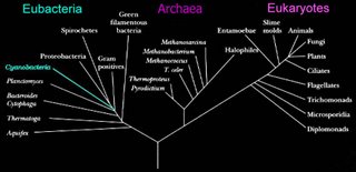
photosynthetic bacteria. The Chlorobiaceae form a coherent group that is phylogenetically isolated from all other microbes, so they are the sole occupants of their phylum (Phylum Chlorobi). There are 5 known genera and 14 species in the family Chlorobiaceae with the type strain being Chlorobium limicola. Phylum Bacteroidetes comprise the most closely related phylum.

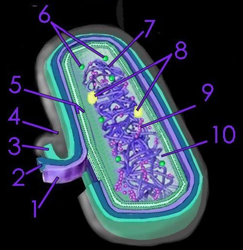
Chlorobiaceae are anaerobic obligate photoautolithotrophs that use sulfide, elemental sulfur or hydrogen as their source of electrons, employing bacteriochlorophylls c, d, and e in vesicles called chlorosomes that are attached to the membrane. As anaerobes, their environment must be oxygen-free, and, as phototrophs, they require light as an energy source. For example, in conducting nonoxygenic photosynthesis employing sulfide ions (H2S) as an electron donor:

CO2 + 2H2S = CH2O + H2O + 2S

The sulfide donates an electron, becoming oxidized and producing globules of elemental sulfur outside the cell, which may then be further oxidized. (By contrast, oxygenic photosynthesis in plants employs water as electron donor and produces oxygen.) Green sulfur bacteria form associations with other microbes that are beneficial both species. When green bacteria convert sulfide to elemental sulfur they store it externally. Some purple bacteria can associate with the green sulfur bacteria and oxidize the sulfur for their own photosynthetic processes. In this relationship, the green sulfur bacteria has a high affinity for sulfide and detoxifies it for the purple bacteria, while the purple bacteria remove the sulfur end product, driving the metabolism of the green sulfur bacteria. There are also examples of green bacteria associating with chemoautolithotrophic bacteria and forming what are called phototrophic consortia in which the green sulfur bacteria form a regular array around a central colorless bacterial cell.

Chlorobiaceae:
(chlorophylls mainly bchl c, d or e)
electron donor for photoautotrophy = S– or So (So globules formed outside cell from S–)
photoheterotrophy? potentially all spp. chemotrophy? none

Due to their requirement for an oxygen-free environment and for sulfide and light, the green sulfur bacteria are restricted to narrow zones in aquatic environments where gradients of light and sulfide overlap under anaerobic conditions. In lakes this narrow zone occurs at depths of 2 to 20 m depending upon the characteristics of the body of water. The Chlorobiaceae also exist in microbial mats, which are communities with different classes of photosynthetic bacteria. Cyanobacteria are often on the top layer, purple sulfur bacteria next and the green sulfur bacteria are on the bottom. This relationship relegates the Chlorobiaceae cells to low light intensities, but minimizes oxygen exposure. The green sulfur bacteria possess highly efficient light-harvesting structures called chlorosomes, which confer their ability to operate at extremely low light intensities .

A species of green sulfur bacteria has been found living near a black smoker off the coast of Mexico at a depth of 2,500 meters beneath the surface of the Pacific Ocean. At this depth, where no sunlight can penetrate, the bacteria, designated GSB1, utilizes the dim glow of the thermal vent.

Green sulfur bacteria are typically nonmotile since only one species is flagellated. Morphologically, green sulfur bacteria are gram-negative rods, vibrio or have a coccus morhphology. Green sulfur bacteria of the genus Prothecocholoris are spherical with a prosthecae emerging from the sphere. Prosthecae have been postulated to be extra
membrane areas that can be filled with additional photosynthetic units. Prosthecae are characteristic appendages that give the cells a star-like shape, and are of a different nature than other external appendages, such as spinae.

green nonsulfur

This item is being cultured.

prochlorophytes

Prochlorophytes : Prochlorococcus Prochloron
chlorophylls chl a and b
electron donor for photoautotrophy = H2O
photoheterotrophy? ? chemotrophy? prob. none

purple sulfur

The purple sulfur bacteria are photosynthetic anaerobic or microaerophilic Proteobacteria found in meromictic lakes and sulfur (hot) springs. These bacteria utilize hydrogen sulfide (H2S) rather than water as their electron donor, so they perform nonoxygenic photosynthesis. (By contrast, oxygenic photosynthesis in plants employs water as the electron donor and produces oxygen.)

Members of the Chromatiaceae family produce internal sulfur granules, while members of the Ectothiorhodospiraceae family produce external sulfur granules on oxidation of H2S:

CO2 + 2H2S → CH2O + H2O + 2S

Meromictic lakes are permanently stratified lakes with denser (typically saline) water at the bottom. Under conditions that support purple sulfur bacteria, sulfide is produced in the bottom sediments and diffuses upward into the anoxic bottom waters, where the purple sulfur bacteria form blooms, usually in association with green phototrophic bacteria.

Sulfur springs possess geochemically or biologically produced hydrogen sulfide, which can trigger the formation of blooms of purple sulfur bacteria.

Nonoxygenic photosynthesis
Purple sulfur families:
Chromatiaceae Order Chromatiales (chlorophylls bchl a or b)
electron donor for photoautotrophy = S– or So or H2 (So globules formed inside cell from S–)
photoheterotrophy? some spp. chemotrophy? some spp.

Ectothiorhodospiraceae (chlorophylls bchl a or b)
electron donor for photoautotrophy = S– or H2 (o globules formed outside cell from S–)
photoheterotrophy? possibly all spp. chemotrophy? some spp.

purple nonsulfur

The purple nonsulfur photosynthetic eubacteria constitute a non-taxonomic group, most of which can grow as photoheterotrophs, photoautotrophs or chemoheterotrophs. Chemotrophic growth for the purple nonsulfur bacteria is achieved by respiration, although some exceptional strains and species can obtain energy by fermentation or anaerobic respiration.

These organisms switch modes depending on environmental conditions, particularly: presence or lack of oxygen, availability of carbon source (CO2 for autotrophic growth, organic compounds for heterotrophic growth), and availability of light. Sulfide can be employed as an electron donor when present at low concentrations. However, higher concentrations of H2S (in which the purple sulfur and green sulfur bacteria can thrive) are toxic to purple nonsulfur bacteria.


There are four phylogenetically distinct groups of purple nonsulfur bacteria:
Order "Rhodocyclales" Family "Rhodocyclaceae" are β-proteobacteria,
Rhodospirillum, Rhodopseudomonas, and Rhodobacter groups are α-proteobacteria.

Purple nonsulfur families: Rhodospirillaceae Rhodospirillum
chlorophylls bchl a or b
electron donor photoautotrophy = H2 probably for all, and for some low levels of S–, S2O3–, So
photoheterotrophy? all spp. chemotrophy? probably all spp.

Rickettsiales

Mitochondria are believed to have developed from an endosymbiotic union with alpha-proteobacteria, specifically the Rickettsiales.

The genome of Rickettsia prowazekii is similar to mitochondrial genomes. Phylogenetic analyses indicate that R. prowazekii is more closely related to mitochondria than is any other microbe yet analyzed. Neither genome contains genes required for anaerobic glycolysis. R. prowazekii does contain a complete set of genes encoding components of the tricarboxylic acid cycle and the respiratory-chain complex, so ATP production in Rickettsia is the same as that in mitochondria. The genes from Rickettsia prowazekii encoding cytochrome b (cob) and cytochrome c oxidase subunit I (cox1) provide further phylogenetic evidence for a link with nitochondrial origins.

Rickettsia are obligate intracellular pathogens. They are dependent on invasion, growth, and replication within the cytoplasm of a eukaryotic host cell. Rickettsial progeny are released to initiate a new infection cycle when the host cell undergoes lysis. It is theorized that Rickettsiae survive and reproduce within host cells because they are capable of "suppression of the antimicrobial activities of the eukaryotic target cells, specifically monocytes/macrophages" (Radulovic et al. 2001).

A phylogenetic analysis of the cytochrome b and cytochrome c oxidase I genes supports an origin of mitochondria from within the Rickettsiaceae.
We have cloned and sequenced the genes encoding cytochrome b (cob) and cytochrome c oxidase subunit I (cox1) from Rickettsia prowazekii, a member of the alpha-proteobacteria. The phylogenetic analysis supports the hypothesis that mitochondria are derived from the alpha-proteobacteria and more specifically from within the Rickettsiaceae. We have estimated that the common ancestor of mitochondria and Rickettsiaceae dates back to more than 1500 million years ago.

Sicheritz-Ponten T, Kurland CG, Andersson SG. A phylogenetic analysis of the cytochrome b and cytochrome c oxidase I genes supports an origin of mitochondria from within the Rickettsiaceae. Biochim Biophys Acta. 1998 Jul 20;1365(3):545-51.

The genome sequence of Rickettsia prowazekii and the origin of mitochondria.
We describe here the complete genome sequence (1,111,523 base pairs) of the obligate intracellular parasite Rickettsia prowazekii, the causative agent of epidemic typhus. This genome contains 834 protein-coding genes. The functional profiles of these genes show similarities to those of mitochondrial genes: no genes required for anaerobic glycolysis are found in either R. prowazekii or mitochondrial genomes, but a complete set of genes encoding components of the tricarboxylic acid cycle and the respiratory-chain complex is found in R. prowazekii. In effect, ATP production in Rickettsia is the same as that in mitochondria. Many genes involved in the biosynthesis and regulation of biosynthesis of amino acids and nucleosides in free-living bacteria are absent from R. prowazekii and mitochondria. Such genes seem to have been replaced by homologues in the nuclear (host) genome. The R. prowazekii genome contains the highest proportion of non-coding DNA (24%) detected so far in a microbial genome. Such non-coding sequences may be degraded remnants of 'neutralized' genes that await elimination from the genome. Phylogenetic analyses indicate that R. prowazekii is more closely related to mitochondria than is any other microbe studied so far.

Andersson SG, Zomorodipour A, Andersson JO, Sicheritz-Ponten T, Alsmark UC, Podowski RM, Naslund AK, Eriksson AS, Winkler HH, Kurland CG. The genome sequence of Rickettsia prowazekii and the origin of mitochondria. Nature. 1998 Nov 12;396(6707):133-40. Comment Nature. 1998 Nov 12;396(6707):109-10.

The genome of Rickettsia prowazekii and some thoughts on the origin of mitochondria and hydrogenosomes.
The sequence of an alpha-proteobacterial genome, that of Rickettsia prowazekii, is a substantial advance in microbial and evolutionary biology. The genome of this obligately aerobic intracellular parasite is small and is apparently still undergoing reduction, reflecting gene losses attributable to its intracellular parasitic lifestyle. Evolutionary analyses of proteins encoded in the genome contain the strongest phylogenetic evidence to date for the view that mitochondria descend from alpha-proteobacteria. Although both Rickettsia and mitochondrial genomes are highly reduced, it appears that genome reduction in these lineages has occurred independently. Rickettsia's genome encodes an ATP-generating machinery that is strikingly similar to that of aerobic mitochondria. But it does not encode homologues for the ATP-producing pathways of anaerobic mitochondria or hydrogenosomes, leaving an important issue regarding the origin and nature of the ancestral mitochondrial symbiont unresolved.

Muller M, Martin W. The genome of Rickettsia prowazekii and some thoughts on the origin of mitochondria and hydrogenosomes. Bioessays. 1999 May;21(5):377-81.

sulfur bacteria

The rather confusing term 'sulfur bacteria' encompasses green sulfur bacteria plus purple sulfur bacteria (distinguised from green nonsulfur and purple nonsulfur bacteria) .

The green sulfur bacteria (Chlorobiaceae) are photosynthetic bacteria that form a coherent group phylogenetically isolated from all other microbes, such that they are the sole occupants of their phylum (Phylum Chlorobi).

The purple sulfur bacteria are photosynthetic anaerobic or microaerophilic Proteobacteria found in meromictic lakes and sulfur (hot) springs.

Chlorobiaceae (green sulfur) are anaerobic obligate photoautolithotrophs that use sulfide, elemental sulfur or hydrogen as their source of electrons, employing bacteriochlorophylls c, d, and e in vesicles called chlorosomes that are attached to the membrane. As anaerobes, their environment must be oxygen-free, and, as phototrophs, they require light as an energy source.

In conducting nonoxygenic photosynthesis, green and purple sulfur bacteria employ sulfide ions (H2S) as an electron donor:

CO2 + 2H2S → CH2O + H2O + 2S

Members of the purple Chromatiaceae family produce internal sulfur granules, while members of the Ectothiorhodospiraceae family produce external sulfur granules on oxidation of H2S.
. . . since 10/06/06
Search 
Search 
for